 The medical specialty known as radiology focuses on diagnosing and treating disease using imaging methods (like X-rays). It can be employed as a diagnostic tool to establish whether a medical condition is present, interventional equipment to perform a procedure, or a treatment tool, for example, to administer radiation therapy to address melanoma.
Varied medical specialists may be involved in a radiological test or operation. Radiologic methods can help treat many disorders. There are also different types of radiology scans & procedures for diagnostic imaging.
Table of Contents
Various Radiology Scans and Procedures
Here is a list of some common types of radiology scans & procedures conducted regularly at radiology clinics.
X-Rays
Plain radiographs or X-rays are frequently used to examine the belly, chest, or bones. Denser components, like bones, look white on an X-ray because they are opaque. On the other hand, air-filled organs appear black, like the lungs. Most of the body’s structures fall between such two on a grey scale.
X-rays can be utilised to identify health problems like fractures, pneumonia, or bowel obstructions. But frequently, more imaging tests are also required.
The effectiveness of X-rays can be restricted based on the region of the body scanned. For example, an anomaly is less likely to be seen on an arm X-ray since many structures overlap. It includes the clavicle, heart, and lungs on the left side of the chest.
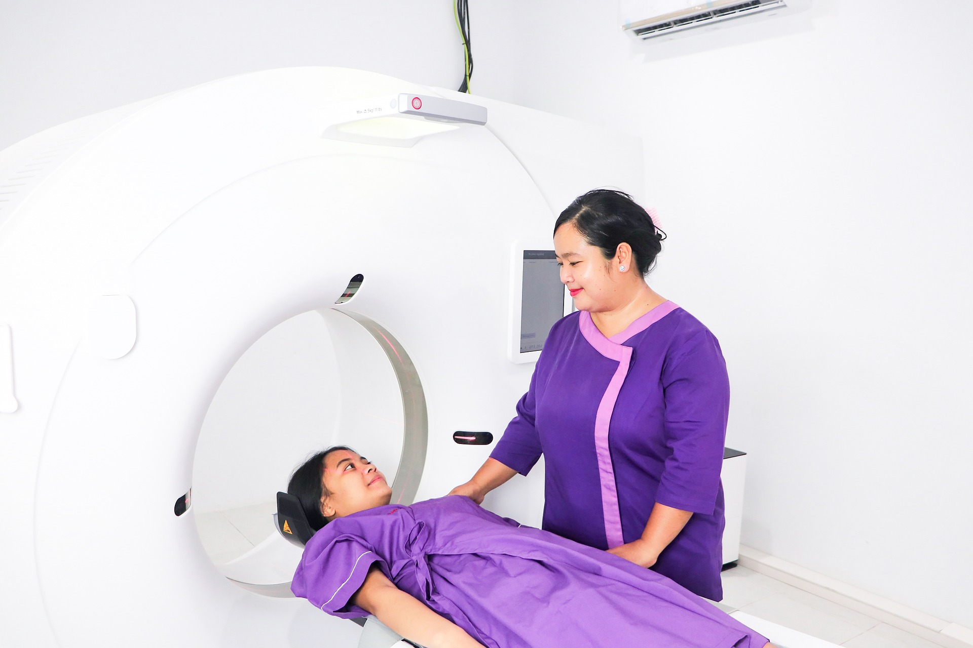
Computerised Tomography (CT)
Using a computer and a sequence of X-rays, computed axial tomography (CAT scans or CT scans) creates a cross-sectional image of the inside of the body. Compared to an X-ray, a CT scan offers more information that can more clearly distinguish places where tissues overlap.
Furthermore, a CT scan helps discover more minor irregularities than an ordinary X-ray. The clarity of specific areas, such as the digestive tract, can be further improved by applying contrast dyes during CT scans. Specific CT treatments, including CT angiography, may offer information in cases that require more medical interventions.
Imaging Using Magnetic Resonance (MRI)
During the magnetic resonance imaging procedure, a radiologist uses powerful electromagnetic fields and radio waves to make an image of the inside of the body.
MRI has made it possible for medical professionals to identify illnesses that, in the past, were only clinically believed to exist. It includes the brain, spinal cord, and brachial plexus abnormalities. For instance, practitioners can now identify multiple sclerosis using an MRI when this diagnosis was only possible based on an evaluation of symptoms.
While MRI is generally a better test for examining soft tissue, such as the nervous system, spinal cord, neurons, muscles, ligaments, and breast tissue, a CT scan is often a better technique for examining bones and arteries. Besides, MRI is a more reliable method of detecting breast cancer than mammography.
Arthrogram
In an arthrogram, a specific contrast material—often referred to as dye—is injected into your body. An X-ray, endoscopic, MRI, or CT scan is then performed.
Arthrograms produce images that are more detailed than tests that lack contrast. Radiologists frequently use these tests to examine joints more closely and determine what is causing pain or functionality loss. An arthrogram uses contrast fluid to let medical professionals see your bones and tissues in greater detail.
An arthrogram also identifies the underlying cause of joint pain or physical disabilities. The examination can detect tears in your joints’ ligaments, muscles, cartilage, and capsules. Additionally, it can look for painful human bones or dislocated joints.
Conclusion
Thus, radiology has a far broader scope than some people realise and isn’t confined to X-rays and CT scans. Interventional radiology, once primarily used to diagnose illnesses and injuries, now offers alternatives to earlier, more intrusive procedures. The best radiologists will suggest the right types of radiology scans & procedures based on the patient’s medical condition.